Wednesday, December 25, 2013
Answer 30
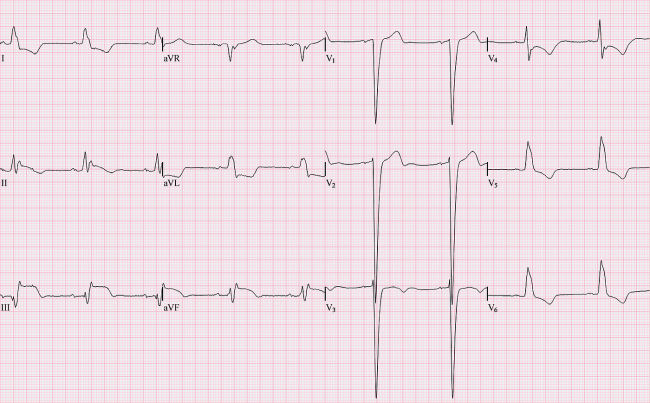
30. Choice A is the correct answer. With right ventricular hypertrophy, there is a large R wave in lead V1. There is a more positive deflection toward the V1 electrode and expect the QRS complex more upright than normal. The S wave in lead V1 is smaller than the R wave. With left ventricular hypertrophy, the left ventricle causes QRS complexes to be bigger in both height and depth in leads V1-V6. There is a larger R wave in lead V5 because it is right over the left ventricle. There is often inversion of the T wave. With atrial hypertrophy there is a biphasic P wave in lead V1. If the initial component is the largest there is right atrial hypertrophy. If the terminal portion of the P wave in lead V1 is the largest, it is left atrial hypertrophy.
Question 30
30. Please identify the abnormality on the EKG listed below:

A. Right Ventricular Hypertrophy
B. Left Ventricular Hypertrophy
C. Left Atrial Hypertrophy
D. Right Atrial Hypertrophy
A. Right Ventricular Hypertrophy
B. Left Ventricular Hypertrophy
C. Left Atrial Hypertrophy
D. Right Atrial Hypertrophy
Answer 29
29. Choice D is the correct answer. Hypocalcemia causes prolonged QT interval. This is best assessed in leads V5 and V6. Hypercalcemia causes shortened QT interval. Hypokalemia causes flattened T waves and U waves. Hyperkalemia causes peaked T waves.
Question 29
29. Please identify the abnormality on the EKG listed below:

A. Hyperkalemia
B. Hypokalemia
C. Hypercalcemia
D. Hypocalcemia
A. Hyperkalemia
B. Hypokalemia
C. Hypercalcemia
D. Hypocalcemia
Answer 28
28. Choice B is the correct answer. Left bundle branch block is demonstrated by the R and R' waves in leads V5 and V6. If it is only present in one lead it is referred to as a incomplete left bundle branch block. Hypokalemia is when the T wave is flattened and there may be a U wave present. This is not ventricular tachycardia.
Question 28
28. Please identify the abnormality on the EKG below:
A. Incomplete Left Bundle Branch Block
B. Left Bundle Branch Block
C. Ventricular Tachycardia
D. Hypokalemia
Tuesday, December 24, 2013
Answer 27
27. Choice B is the correct answer. It is a narrow complex tachycardia. It is a rapid sudden onset of a heart rate originating from an ectopic foci form the AV junction. The P wave is inverted and occurring before during or after the QRS complex. Rate is between 150-250 beats per minute. PAT is when the ectopic focus occurs from the atria. Often it is hard to tell if the the P wave is present or not. Collectively these tachycardias are referred to as supraventricular tachycardia. Ventricular tachycardia is a wide complex tachycardia that has a rate over 150 beats per minute. Atrial Fibrillation is an irregularly irregular rhythm that the ectopic focus occurs from the atria.
Subscribe to:
Posts (Atom)